Case Report
The efficacy of complex Decongestive Physiotherapy in patients with Bilateral Primary Lower Extremity Lymphedema and Untreatable multiple health conditions: A Case Report

Hümeyra Kiloatar PT*
Department of Physiotherapy and Rehabilitation, School of Health Science, Dumlupinar University, Campus of Evliya Celebi, 43100 Kutahya, Turkey
*Address for Correspondence: Hümeyra Kiloatar PT, Department of Physiotherapy and Rehabilitation, School of Health Science, Dumlupinar University, Campus of Evliya Celebi, 43100 Kutahya, Turkey, Tel: +(90)05357690421; Email: [email protected]
Dates: Submitted: 10 August 2017; Approved: 07 September 2017; Published: 08 September 2017
How to cite this article: Hümeyra Kiloatar PT. The efficacy of complex Decongestive Physiotherapy in patients with Bilateral Primary Lower Extremity Lymphedema and Untreatable multiple health conditions: A Case Report. J Nov Physiother Rehabil. 2017; 1: 093-098.
DOI: 10.29328/journal.jnpr.1001011
Copyright License: © 2017 Hümeyra Kiloatar PT. This is an open access article distributed under the Creative Commons Attribution License, which permits unrestricted use, distribution, and reproduction in any medium, provided the original work is properly cited.
Abstract
Background: Primary lymphedema occurs as a result of genetic abnormalities of the lymph system. Currently, complex decongestive therapy is accepted as the standard treatment of the lymphedema. In this case presentation, we described the management of bilateral primary lower extremity lymphedema and the use of complex decongestive therapy.
Case Report: A 62 years old female patient had stage III primary lymphedema on her left lower extremity and stage II primary lymphedema on her right lower extremity. The patient, who had morbid obesity, also had untreatable sleep apnea, urinary incontinence, umbilical hernia and hypertension controlled by drugs. She had stage 4 gonarthrosis according to Kellgren – Lawrence classification in her both knees. The patient received complex decongestive therapy as an outpatient.
After 27 sessions of complex decongestive therapy, edema reduced in both lower extremities. Before the treatment started, the patient couldn’t go up and down stairs, get out and had difficulty mobility in the home. But after the treatment, the patient could go up and down 16 stairs by holding the railing, get out by two walking sticks and had less difficulty mobility in the home. However, due to gonarthrosis in her knees, her pain did not diminish.
Conclusion: Complex decongestive therapy is effective in the management of bilateral primary lower extremity lymphedema, which progressed with multiple health conditions.
Introduction
Lymphedema, which occurs as a result of congestion of protein-rich fluid within the lymphatic system, is a chronic, progressive, disabling, and incurable but treatable health condition [1]. Lymphedema is classified as primary and secondary lymphedema according to its etiology [2]. Primary lymphedema occurs as a result of genetic abnormalities of the lymphatic system [2].
The diagnosis of lymphedema is based on the full and complete history and physical exam [3]. Current and past medical history (traumatic events, surgery, cancer diagnosis, cancer treatment with radiation, cardiac issues, venous insufficiency, and a family history of lymphedema) should be noted.
Extensive visual assessment and palpation of the skin is performed evaluating the texture, pitting status, stemmer’s sign, fibrosis, color, temperature, deepening of the skin folds, nail quality, hyperkeratosis, dermatitis, ulceration, cyst status and the presence of papillomas in the affected extremity [4]. Assessment of the edema must be performed in the involved as well as the uninvolved extremities and proper documentation is essential. The two ways of accomplishing this are volumetrics and circumferential measurements [5]. Digital photography should be performed [6].
Currently, complex decongestive therapy is accepted as the standard treatment of the lymphedema [7].
We described the management of bilateral primary lower extremity lymphedema and the use of complex decongestive therapy.
Case Report
Patient history
The patient was a 62 years old female. She was 160 cm height, and 125 kg weight. Lymphedema started in her left extremity in 2006 without obvious reason and appeared in both extremities in 2015 (Figure 1). She was diagnosed with lymphedema in 2015 but due to living in a rural area, she could not find physiotherapist who have education about lymphedema and complex decongestive therapy. In the lymphoscintigraphy, which was taken in 2015, normal lymphatic drainage in the right lower extremity, slow lymphatic flow and partial obstruction in the left lower extremity have been reported. Dilatation and reflux in left great saphenous vein have been reported in bilateral venous Doppler ultrasound examination. Additionally, edema has been detected in both lower extremities. Based on the results of lymphoscintigraphy and ultrasound examination, using pneumatic compression device has been considered and she has used pneumatic compression device at home for the last 2 years.
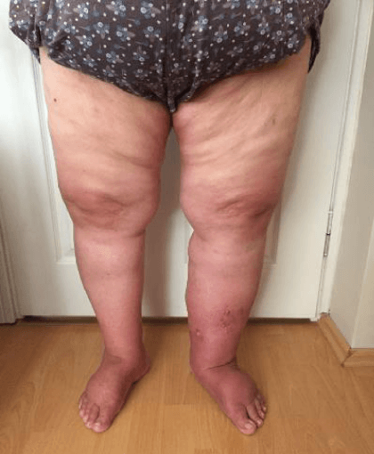
Figure 1: The patient’s right and left lower extremities before the treatment.
Diagnosis
According to clinical examination was done by physiotherapist, the patient was diagnosed. There were non-pitting edema, fibrosis, and positive Stemmer’s sign. Tissue density of the left extremity was harder. A lot of cysts were seen on the medial aspect of her left foot. The cyst was filled with chyle, which is a fluid abound with lipids (Figure 2). With these findings, it was decided that the left extremity had Stage III lymphedema and the right extremity had Stage II lymphedema. When the age of onset of the disease is considered, it can be said that it is lymphedema tarda.
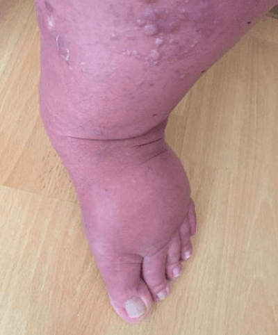
Figure 2: Chyli cysts on the left extremity.
Examination
The patient was mobile at home and was walking with two walking sticks. She had stage 4 gonarthrosis according to Kellgren-Lawrence classification in her both knees. The patient had morbid obesity. She also had untreatable sleep apnea, urinary incontinence, umbilical hernia, and hypertension controlled by drugs. Her family history included lymphedema. She was using Zaldiar (1*2), Fludex (1*1), Coraspirin (1*1), Venorutan(1*2), and Co-diovan (1*1) daily. During the initial examination, the patient’s pain, circumference measurement, and mobility were evaluated and images were taken.
Edema assessment
Edema was evaluated by measuring the extremity circumference. The measurements were made before and after the treatment at the level of metatarsal heads, 4 cm proximal to metatarsal heads, at the level of the lateral malleolus, and with 4 cm intervals thereafter. The measurements are given in table 1.
| Table 1: Before and after treatment the extremity circumference measurements. | ||||||
| RIGHT (cm) | LEFT (cm) | |||||
| BEFORE | AFTER | DIFFERENCE | BEFORE | AFTER | DIFFERENCE | |
| Metatarsal head | 24 | 23,5 | 1,5 | 27 | 23,5 | 3,5 |
| 5 cm above | 27 | 24,5 | 2,5 | 28 | 25 | 3 |
| Lateral malleol | 29 | 29 | 0 | 34,5 | 32,5 | 2 |
| 4 cm above | 28 | 26,5 | 1,5 | 34,5 | 29 | 5,5 |
| 8 cm above | 30,5 | 28,5 | 2 | 38,5 | 32 | 6,5 |
| 12 cm above | 34 | 33 | 1 | 43,5 | 36,5 | 7 |
| 16 cm above | 39 | 37 | 2 | 48 | 42 | 6 |
| 20 cm above | 43 | 42,5 | 1,5 | 50,5 | 46 | 4,5 |
| 24 cm above | 45,5 | 44 | 1,5 | 51 | 49 | 2 |
| 28 cm above | 45 | 43 | 2 | 49,5 | 47 | 2,5 |
| 32 cm above | 44 | 44 | 0 | 46 | 45 | 1 |
| 36 cm above | 53 | 53 | 0 | 54 | 52 | 2 |
| 40 cm above | 58,5 | 58 | 0,5 | 59,5 | 58,5 | 1 |
| 44 cm above | 61 | 61 | 0 | 63,5 | 61 | 2,5 |
| 48 cm above | 66,5 | 65,5 | 1 | 67 | 65 | 2 |
| cm: centimeter | ||||||
Pain
The pain score was 10 according to visual analog scale (VAS) in both knees. The pain could be related to gonarthrosis and immobility. Accordingly VAS, score of 0 refers to ‘no pain’ and score of 10 calls‘worst imaginable pain’.
Interventions
The patient was managed as an outpatient because the patient could not go up and down the stairs and there was not an elevator in her house. At first, the patient was informed about her disease, treatment, and her and her family’s responsibilities. We encouraged the patient to participate in the treatment. The patient was taken into treatment for 6 days in a week.
Manual lymph drainage
The session was started with the manual lymph drainage performed by a physiotherapist. Simultaneously, manual lymph drainage was taught to the patient. Since the body edema was too much, lower body drainage was performed primarily for 5 sessions. The session was ended with the application of short stretch bandages to both lower extremities. Then, the sessions continued with left lower extremity since the edema was more significant on the left. The treatment continued with twelve sessions of left extremity drainage and simultaneous 6 sessions of lower body drainage. Because the patient had umbilical hernia, abdominal techniques weren’t applied. Abdominal techniques are contraindication for umbilical hernia. Only deep breathing techniques were applied at beginning of the manuel lymph drainage. After drainage, both lower extremities were bandaged. Lastly, 10 sessions of right extremity drainage were applied.
Skin care
After manual lymph drainage, skin care was performed with a moisture cream Sebamed® with a pH value of 5.5 every day. After the 4th session, the patient’s cysts ruptured and a heavy exudation occurred. During this period, the wound was cleaned with physiologic saline and dressed with sterile gauze. Compression bandaging of both extremities continued.
Multilayer bandaging
After manual lymph drainage and skin care, compression was performed by a physiotherapist with short stretch bandages. At first, to protect the skin, stockinette was employed by keeping the toes open. The lower extremity was bandaged with a 100% polyurethane pad by forming a cylinder up to the thigh. In order to prevent the cyst from rupturing, and protect the wound occurred after spontaneous rupture of the cyst, a U shape was made with 10 mm thick pad and placed under the bandage. Then, bandaging was made with 6 cm, 8 cm, 10 cm, and 12 cm short stretch bandages.
Exercise
After bandaging, an active assisted range of motion exercise with 10 repeats was performed for ankle, knee, and hip joints. Walking training was given with walker to facilite walking. Phase II education was given to the patient simultaneously with Phase I. Self-drainage, self-bandaging, skin care and exercises were taught. The things that the patient should not do were explained and given in written.
The post treatment instructions are in the following section:
1. You are careful that your clothes are enough large size to leave no mark on the skin.
2. You don’t opt for a hot and tropical region for your holiday.
3. You keep away from sauna, bath and spa.
4. You don’t smoke.
5. You relax at regular intervals during exercise to avoid pain in your extremity.
6. You do exercise with your pressure garment.
7. When you travel by plane, you must wear pressure garment.
8. If you notice signs of infection in your body, you consult your doctor immediately.
Pressure garment
Considering that the patient was immobile, the patient did not have anyone to help her, and the severity of the disease, JOBST® class III compression garment was decided. The pressure of class III garment is 34-46 mmHg. Since the patient had incontinence, an AG style compression garment was preferred rather than a pantyhose style. A foot cap was added to compression garment since there was significant edema over the foot. The patient was informed about using the compression garment (Figure 3).
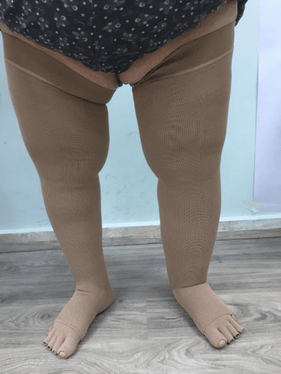
Figure 3: The patient’s right and left lower extremities with pressure garments after the treatment.
Results
After 27 sessions of complex decongestive therapy, edema was reduced in both lower extremities of the patient (Table 1). Before the treatment started, the patient couldn’t go up and down stairs, get out and had difficulty mobility in the home. But after the treatment, the patient could go up and down 16 stairs by holding the railing, get out by two walking sticks and had less difficulty mobility in the home. She started to move more easily in her bed. The patient stated that she felt herself better. Her pain did not diminish. Accorging to VAS, the pain score was still 10. Because the pain didn’t associated lymphedema [4], probably because of gonartrosis. The good outcomes are maintained after CPT at 12 months follow-up.
Discussion
Lymphedema is a chronic, progressive disease that limits the patient’s independence. If not treated, the disease will progress and cause disability in time [8]. There is a lack of knowledge regarding the management of lymphedema among health professionals around the world [9]. Since health professionals are not aware of the importance of early diagnosis and treatment, patients are often neglected in the early stages [10]. Holtgrefe applied complex decongestive physiotherapy for 8 weeks, 2 sessions in each, in a patient with stage II bilateral secondary lower extremity lymphedema. At the end of the treatment, 9% improvement on the left extremity and 10% improvement on the right extremity were observed. Before the treatment, the patient had the pain which score was 10 according to VAS. But after the treatment, the pain score was 0 [8].
Combined microsurgical treatment and physical treatment of lymphedema is becoming a new standard in the treatment of lymphedema. Patients who demonstrate responsiveness to an adequate trial of conservative therapy and early stages of lymphedema are favorable candidates for microsurgical treatment [1,11]. Patients undergoing microsurgical lymphatic procedures are immediately prescribed continuous compression garment use postoperatively, which makes definitive conclusions regarding a direct benefit from the surgical procedure above the improvement from compression therapy difficult [1]. Microsurgical operations can restore lymphatic drainage, both in the short- and long-term, and the best results are obtained when these surgical procedures are combined with physical rehabilitation methods [12]. But, all current therapies, none of the available surgical techniques completely address patients’ underlying lymphatic dysfunction, although patients can achieve resolution of their symptoms and extremity swelling with sustained adherence to compression garment regimens postoperatively [1].
However lymphoedema management and basic principles of treatment are used in developed countries, it can’t be transmitted developing countries due to financial limitations in the materials for treatment, lack of health care providers and lack of information about lymphoedema management. In our case, phase I of complex decongestive therapy, which was applied for 27 sessions, reduced patient’s edema, improved the patient’s mobility level and restored the patient’s self-confidence. Despite her edema reduced, she continued to use walking sticks. Gonarthrosis on the patient’s knees limits her independence. Umbilical hernia, sleep apnea, and primary lymphedema hinder the patient from undergoing knee replacement surgery. If a multidisciplinary team can be formed, knee replacement surgery can be planned for the patient in the future. Lymphedema, which can occur after a knee replacement surgery, will be followed. Complex decongestive therapy is effective in the management of progressed bilateral primary lower extremity lymphedema which worsened due to multiple health conditions.
References
- Warren AG, Brorson H, Borud LJ, Slavin SA. Lymphedema: a comprehensive review. Ann Plast Surg. 2007; 59: 464-472. Ref.: https://goo.gl/pSGEyS
- Kerchner K, Fleischer A, Yosipovitch G. Lower extremity lymphedema: Update: Pathophysiology, diagnosis, and treatment guidelines. J Am Acad Dermatol. 2008; 59: 324-331. Ref.: https://goo.gl/Y99FWU
- International Society of Lymphology. The diagnosis and treatment of peripheral lymphedema: 2013 Consensus Document of the International Society of Lymphology. Lymphology. 2013; 46: 1-11. Ref.: https://goo.gl/Ey65To
- Gary DE. Lymphedema diagnosis and management. J Am Acad Nurse Pract. 2007; 19: 72-78. Ref.: https://goo.gl/r5M1hs
- Zuther J. Lymphedema management: The comprehensive guide for practitioners. Thime. 2009.
- Lawenda BD, Mondry TE, Johnstone PA. Lymphedema: a primer on the identification and management of a chronic condition in oncologic treatment. CA Cancer J Clin. 2009; 59: 8-24. Ref.: https://goo.gl/gU4pPZ
- Bogan LK, Powell JM, Dudgeon BJ. Experiences of living with non-cancer-related lymphedema: Implications for clinical practice. Qual Health Res. 2007; 17: 213-224. Ref.: https://goo.gl/HPK1n7
- Holtgrefe KM. Twice-weekly complete decongestive physical therapy in the management of secondary lymphedema of the lower extremities. Phys Ther. 2006; 86: 1128-1136. Ref.: https://goo.gl/p8Ue2y
- Bland KL, Perczyk R, Du W, Rymal C, Koppolu P, et al. Can a practicing surgeon detect early lymphedema reliably? Am J Surg. 2003; 186: 509-513. Ref.: https://goo.gl/pUUcKM
- Stout NL, Brantus P, Moffatt C. Lymphoedema management: an international intersect between developed and developing countries. Similarities, differences and challenges. Glob Public Health. 2012; 7: 107-123. Ref.: https://goo.gl/A34fVH
- Kung TA, Champaneria MC, Maki JH, Neligan PC. Current concepts in the surgical management of lymphedema. Plast Reconstr Surg. 2017; 139: 1003-1013. Ref.: https://goo.gl/GR2GPk
- Boccardo F, Fulcheri E, Villa G, Molinari L, Campisi C, et al. Lymphatic microsurgery to treat lymphedema: techniques and indications for better results. Ann Plast Surg. 2013; 71: 191-195. Ref.: https://goo.gl/jhE6o4